Voiceover: This podcast is for informational and educational purposes only. It is not medical care or advice. Clinicians should rely on their own medical judgements when advising their patients. Patients in need of medical care should consult their personal care provider. Welcome to "That's Pediatrics", where we sit down with physicians, scientists, and experts to discuss the latest discoveries and innovations in pediatric healthcare.
Dr. Allison “Alli” Williams: I'm Alli Williams. I'm one of the hospitalists here at UPMC Children's Hospital of Pittsburgh.
Dr. Sameer Agnihotri: I'm Sameer Agnihotri. I'm one of the scientists here at Children's Hospital.
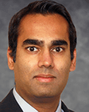
Dr. Williams: And we are so excited today to be joined by Dr. Laura Olivieri. She is one of our cardiologists who also specializes in cardiac imaging. Thank you so much for being here. Can you describe a little bit about what brought you to the Pittsburgh area?
Dr. Laura Olivieri: Yeah, it's a great question. So I actually spent a lot of time in... I didn't grow up in Pittsburgh, but I spent a lot of time in Pittsburgh when I was little. Actually all through my childhood, because my mom grew up here and my grandparents were here. They lived in Bellevue.
Dr. Williams: Oh, nice.
Dr. Olivieri: Yeah. And so, name a major holiday in the eighties and nineties. I was probably here. So I've always honestly, really enjoyed spending time in Pittsburgh for obvious reasons. But I've always felt like really comfortable here. Long story long, I guess, my parents moved back to the Pittsburgh area, and so when my family and I were preparing for the next career move, Pittsburgh was not just a familiar and friendly place to be, but the combination of the culture here and the outstanding outcomes in cardiology and cardiac surgery were really attractive to me to make my next move.
Dr. Agnihotri: Wow, that's fantastic. Can you tell us a little bit about your training and how you got to your career job here?
Dr. Olivieri: To be totally honest, I guess it starts when I was a kid. I don't know if you want to go back that far or not.
Dr. Williams: But as far as you want.
Dr. Agnihotri: As far as you want.
Dr. Olivieri: As far as as I want. Okay. So my brother was born with a congenital heart defect. One in a hundred babies in the world is born with one. My brother was one of them. He was born with total anomalous pulmonary Venus return and needed a life-saving surgery when he was little. And so from the time I was little, he's my little brother, I kind of was interested and kind of geared towards medicine and I guess specifically pediatric medicine. So I went to college. I did my undergrad in engineering, but then went to medical school in Chicago and did my pediatric training at Brown in Rhode Island, which was lovely. And then went to DC and was in DC for 15 years for pediatric cardiology fellowship. I trained in imaging there. And then that was where I started my career.
Had had a great time, loved the program there, and loved living in DC. It was great. I learned a lot there and I'm really indebted to a lot of people there for teaching me things and pointing out different interesting things. And that's honestly where I fell in love with imaging as well. So I guess in what I do here is I direct non-invasive cardiac imaging. So I do echo CT and MR. And although I don't do fetal echocardiography, I support the people who do fetal echocardiography, which is an important part of our program. So that's kind of been my path.
Dr. Agnihotri: Amazing.
Dr. Williams: Having an engineering background seems unusual for medicine, but also probably quite applicable when it comes to the heart. I feel like the heart is an own machine in and of itself.
Dr. Olivieri: Yeah, absolutely. And I think within cardiology, I feel like it's probably not that rare to find people in cardiology who also have engineering backgrounds. And I don't know, I don't want to overstate my engineering background.
Undergrad was a long time ago. Yeah. Calculators had barely been invented. No, I'm kidding. We had Abacus. No. And I think this goes for all of us, is you kind of learn your patterns of how you think about the world and analyze the world when you're in that college age. And so I think learning to think about things a little bit like an engineer was super helpful for medicine. And I think it was nice, and I don't know what you all think, but I think it was nice to have a little bit of a background in something, not medicine, and then go to medicine. I don't know if you found the same to be true.
Dr. Williams: I think that that's wonderful. I also know that in cardiac imaging, there's been... At least I feel like as a hospitalist, that there's been a lot of changes over the past few years with it and a lot of expansion. Could you kind of comment on maybe one of your passion projects related to the imaging in pediatric cardiac imaging?
Dr. Olivieri: Yeah, yeah, I absolutely agree with you. There really has been a big shift in our field. So one of the big shifts, it's funny, on my way down here to talk to you I was just thinking about this. When my brother was born in 1983, the reason they found out he had a congenital heart defect was because the physicians taking care of him, performed a cardiac catheterization, so did essentially invasive cardiac imaging and injected contrast, and did fluoroscopy used radiation sedation, contrast all this exposure to get an understanding of what was wrong so it could be surgical surgically corrected. And probably in the past, I don't know, year or year and a half, I've probably diagnosed two or three other babies with what he had in a completely different way. And I was just thinking about that. I'm like, well, there really is progress.
So now we try to use more. Obviously there's a saying that started in the pediatric radiology community. It's this concept called “image gently,” which I am a total believer in. I think most pediatric imagers are – radiologists and cardiologist. Which is, just use what you need, just use the time that you need and the sedation you need and the radiation exposure if you need it, that you need. Really try and use the technology and tailor your approach to the child and what they need, and only use what you need. Be a bit sparing about these resources and more importantly, these exposures for these babies and children. So I think when we think about the evolution of cardiac imaging, we use ultrasound a lot. Ultrasound's great. It's portable, it's cheap, it's quick, it's fast. It doesn't typically involve any sedation, radiation babies or kids don't need IVs.
It's lovely and it works really well on babies. But as babies grow into little kids and little kids grow into big kids and teenagers, it works a little less well with every year that goes by. And sometimes you just really need to see inside a little bit more clearly. And so cardiac MRI has become a huge technology that's been harnessed around the world really. And in particular in the last few years here by my colleagues, Dr. Adam Christopher and Dr. Tarek Alsaied. They've built a beautiful program here with the support of Dr. Ashak Penagra, providing machine upgrades and really extending the capabilities. So now we can watch blood flowing around in real time without having to do anything to the child to see where do their pulmonary veins go? Is there a narrowing in the aorta or in one of the pulmonary artery? It's beautiful. And in some cases, we don't even need sedation or IVs for contrast.
Dr. Williams: That's amazing.
Dr. Olivieri: Yeah. And again, no radiation exposure with MR, which is great. But you get this beautiful, highly resolved cross-sectional imaging. I guess I'm well suited for my job because I think it's a treat. I just love doing it and looking at it. And I think there's just so much data that's encoded in there that provides a really nice and deep understanding of what the defect is and what we have to do about that next. And then cardiac CT is really exciting as well. Again, it involves a bit of contrast, a bit of radiation exposure for these people that need it. But again, the imaging is beautiful and it leaves nothing. There's no mysteries. It's a very definitive imaging technology that is very useful for people.
So between those three, echo, MR and CT, we definitely try and use them thoughtfully and in a logical way, depending on what the different children have, what problems the children have with their hearts. And I think in particular, MRI really is a place where I guess I personally have put a lot of effort into trying to develop this technology, not just for my use, but for use in the field, trying to do some research on how to make it faster and make it more robust so we don't need sedation or anesthesia for kids in the scanner to make it quicker. And just more, I guess, more reliable.
Dr. Agnihotri: Wow. So speaking to your MRI background, just have to say it is artificial intelligence or computational power, is any of this stuff, these buzzwords we routinely hear, is this helping you? Or can it be harnessed to help you guys?
Dr. Olivieri: That's a great question. So it is helping in some small ways now for sure. And actually, that's a great question because you can imagine, and this is the common theme for us in pediatrics, which is that sometimes adult medicine gets all the cool stuff, and we have to kind of adapt.
Dr. Williams: We're second best that can fiddle, it happens later, all those things, right?
Dr. Olivieri: Little do they realize we're the best.
Dr. Williams: I know. You don't have to tell me.
Dr. Olivieri: So our problems are more complicated, especially in congenital heart disease, where these beautiful little babies and children are just running around with missing sections of their heart. And they could not be happier just running around beautiful little kids that they are. So the point that I'm trying to make, I guess, is that with artificial intelligence, a lot of the algorithms have been designed to recognize big adult hearts with four chambers and crossing outflows typically formed hearts. And then when we try and use those steam algorithms on the children, we take care of, the algorithms are like, no, done. No, I can't do it. Or it tries and it's laughable.
But the nice thing is, over the last probably three years or so, some of these artificial intelligence algorithms have been actually helpful. Because some people, colleagues in congenital heart disease have been able to work, I think, in a more effective way. Both with imaging scientists who create these algorithms and can design the algorithms, I guess, that are needed for imaging analysis, but also work with vendors and companies to try and get these much needed applications in congenital heart disease and pediatric heart disease into their own systems.
Because we have for sure, I think the adult cardiology problems are complicated and adults need cardiac care as well. But the problems that we see and deal with are much more complicated and from an atomic standpoint. And so we really do need more artificial intelligence development. But anyway, I think we're at the start of a very exciting time for artificial intelligence. I know some people in imaging are a little afraid of it, oh, will it take my job? I don't think that's going to happen at all. I think it's going to extend. I really believe it's going to... And knowing people in this field, I really think it's going to extend our capabilities and allow me and my colleagues to do what we do quicker, more reliably, faster, and be able to hopefully extend care to more children. That's the ultimate goal. So yeah, I think absolutely very excited about artificial intelligence and imaging.
Dr. Williams: That sounds amazing. Also, I agree. A little scary. Every time I hear artificial intelligence, I just think of aliens. And I know that's not what it is. It's just where my brain goes when I think about it. This is probably why I'm a pediatrician.
Dr. Olivieri: Totally.
Dr. Williams: That I think about aliens and things like that. Knowing your backstory now about your brother, which we're so appreciative of you sharing with us. Because a lot of us in medicine have personal interests in the fields that we choose. And you talked about the difference of echocardiogram versus MRI. How is MRI being used in the neonatal group? Has it changed what your brother would've experienced at this point in time?
Dr. Olivieri: It's a great question. So the short answer is it's a bit institutionally dependent. So for sure, MR or CT definitely would've changed. Definitely would've changed it. I think every institution's a little bit differently about how they invest in their cross-sectional imaging. So some are more CT heavy, some are more MR Heavy. And for sure at some institutions, if my brother was born in X, Y, or Z place now, yeah, definitely. He could have had an MRI, figured it out and gone to the operating room in maybe a different timeframe. And in other institutions it would've been CT. But for sure, the imaging part of it is, I don't know, I think it represents just a nice development, a nice patient-centric development for the patient experience and for the risk that children undertaken in trying to sort out their diagnoses.
Dr. Williams: And with all of these changes that we've seen in the last 20... Well, I guess if I'm doing math correctly, it's 30 years or 40. Oh, wow. 40 '83, you said. 40. I can do math, I promise. With all the changes we've seen in the last couple of decades with cardiac imaging, where do you see cardiac imaging going from here in the future?
Dr. Olivieri: Oh, this is another great question. I think about this all the time. So the immediate future for sure, there's a really exciting... I don't want to use the word disruptive, but it's a little bit disruptive. Change in CT technology just happened. There's these newer generation scanners. I had nothing to do with the development, but I'm just an admirer of them called photon counting CTs, which drastically... The dose of radiation that someone receives with one of these scanners is drastically reduced with a really lovely and advantageous change in the spatial and temporal resolution. So how long it takes it take the picture, and how resolved crisp the picture is.
So for sure, the CT technology, I think just had a major shift. And I don't know what additional shifts are going to be in the pipeline there, but that was just a major shift. And then in MR Technology, what we're all trying to do, I don't know if either of you has ever had an MRI, it takes a long time, or for sure, you've ordered them for your patients-
Dr. Williams: We've ordered them all the time, and it took a while.
Dr. Olivieri: You may use them in the lab. I don't know.
Dr. Agnihotri: I was a control for a brain MRI back when I was a grad student, and you had to stay really still. But I got to keep the images, and I thought it was so cool.
Dr. Williams: But time-consuming.
Dr. Agnihotri: I do have evidence that I have a brain.
Dr. Olivieri: Very nice. I love that. Actually, when I graduate fourth year fellows, both in DC and then I haven't graduated one yet here, but I'll be sure... Well, since this is a podcast, you can hold me accountable. I would create images of that. I'd be like, oh, just get in the scanner for a second. Create little images of them for their little graduation gifts.
Dr. Agnihotri: I think it's amazing.
Dr. Williams: That is awesome.
Dr. Olivieri: No, I think that's really cool that you have a picture of your brain. I think it's a really neat thing. I think the future of MRI is going to be in much faster acquisition, hopefully. And I think brain MRIs are, this already started, and cardiac MRIs are lagging behind, but hopefully we'll make up the ground. And then the other interesting thing that's going on right now in cardiac MRI, we'll see how this pans out.
We'll see how this ages, I guess, is the advent of low field imaging. So using lower field, so you can take the advantage of lower magnetic field imaging. Is not as restrictive of an environment. And you can take more patients with more devices into the magnet. Confers, a little bit more safety, I guess, and a little bit more of a relaxed environment, rather. Not saying safety isn't important, it just has different standards for what can be brought in and out, which is nice. And then the other thing, which will be coming to children's soon, which is very exciting, is this concept of guiding cardiac catheterization by MRI imaging. Ultra-fast, quickly acquired, acquired fast enough so that it can be acquired and displayed as a person is manipulating a catheter in the heart, which is pretty cool.
So MR guided cardiac catheterization is another interesting, I think an important concept in the development of the cardiac MR field, which is cool. And then the last piece probably that we'll see, I hope, is more artificial intelligence and more images pop up with analysis, with predictions, with prognosis, and imaging analysis pops up with maybe reporting, aligned elements, which would be really cool. This is getting a little, I guess, nerdy, but for sure, I think the technology exists to be able to do that. And I think it'll improve the reliability of the imaging reports, as well as the accessibility to patients. So yeah.
Dr. Agnihotri: Speaking on that, can you highlight a little bit of the research that you're involved with and how that could be incorporated into your clinical practice one day or some of the major questions that are just have not been answered?
Dr. Olivieri: Yeah, that's a great question. So one of the concepts that my colleagues and I are working on, which I think is really exciting, is trying to figure out if we can help the performance and durability of surgical repairs for congenital heart disease in babies in young children, and help them perform well and last into adolescence and adulthood. And one of the ways we're trying to think about this is by obtaining cardiac MRI or cardiac CT as a baby, kind of getting a personalized and special precision look at that child’s specific anatomy, designing a graft or patch or whatever is needed for that baby’s surgery at that time. Designing a very specialized specific patch or graft for them that's 3D printed out of bioresorbable materials, and then would be incorporated and then grow with the baby as the baby becomes a little kid, big kid, and adult. Right now with a lot of this surgical materials that are used in that are lifesaving surgeries and very necessary, and it's a miracle that we have these to use right now, but a lot of the materials that go in with babies and little kids don't last forever. They need to be replaced, which means usually another surgery, at least more procedures and probably another surgery eventually. So that's one of the areas that... I'm not a surgeon, and I'm definitely not like a biomaterials person, but the creation of these would hinge on imaging. And so that's my involvement in that. Which would be cool. That would be really neat.
And then the other thing I think we're doing, I think there's a lot more data specifically in cardiac MRI, a lot more kind of coded 3D and 4D data as to how the ventricles squeeze and the blood flows through the heart that I think we can gain a lot more insights from that than we are doing currently.
And so part of the other project that, again, it's a team sport. It's not just me, but along with, we have an imaging cardiologist and then imaging scientists, together we're trying to figure out if we can maybe create new insights into what the actual hemodynamic problems are and maybe more surrounding timing of the next surgery or timing of the next intervention to understand if we can create an imaging biomarker that might signal, okay, well, when this happens, it's definitely time to do the next surgery or whatever, rather than relying on maybe a less perfect predictor of right of timing. Because obviously surgery's a big deal and you want to time it well and get the best results from it that you can.
Dr. Williams: Yeah, it's the same mentality as the image gently, I believe you described it as, you want to surgery gently. No, that's quite a bad term for it. But you want to only do surgery when you need to. You want to the least amount you can for the safety of the child, but do it at the best time for them too, so it'll last the longest.
Dr. Agnihotri: Exactly. And I love the concept of these biomarkers that you were talking about or imaging markers.
Dr. Olivieri: Yeah. Hopefully. And I think we've already been moderately successful at advancing our understanding of the impact of some of the congenital heart disease, but hopefully there's more to come.
Dr. Williams: And I think there's so much more to come, because if I am not mistaken, UPMC Children's Hospital of Pittsburgh is having an expansion. Which part of this is the Heart Institute? Did I use the right name for that?
Dr. Olivieri: Yes.
Dr. Williams: You can edit that part out. Okay, good. Part of that is the Heart Institute, which I assume will incorporate not only clinical care, but a lot of these imaging modalities that you're talking about that we can provide better care for our community here.
Dr. Olivieri: Yeah, absolutely. We are all in the Heart Institute, Dr. Victor Morrell, Dr. Jackie Kreutzer, Dr. Brian Goldstein, Dr. Adam Christopher, and I, and all the entire team really, we are all thrilled about this. Really, it just represents just a really important investment in the children that we're all taking care of and the families that we're all taking care of to be able to consolidate and streamline their care, their experience, and bring us all together in a physical space. I know it sounds a bit touchy-feely, but it really is important and it's really an impressive investment. I'm so glad to be just a small part of it, looking at things from an imaging perspective, but really happy to be working with that whole team and the team of people who are helping design and do the construction and make sure the building is just outstanding in every way. It's really exciting. We really cannot be more thrilled with that investment.
Dr. Williams: Well, we are so excited to have met you and to learn all about your background and your experiences and are honored to have you here in the Pittsburgh area, to keep improving the care that we provide for children so that it can be even better than it already is under your division's care. So thank you for that.
Dr. Agnihotri: Thank you so much.
Dr. Olivieri: Yeah, thank you so much for having me. This was great.
Dr. Williams: And thanks for listening to That's Pediatrics.
Voiceover: You can find other episodes of That's Pediatrics on Apple Podcasts, Google Podcasts, Spotify, and YouTube. For more information about this podcast or our guests, please visit chp.edu/ThatsPediatrics. If you've enjoyed this episode, please be sure to rate, review and subscribe to keep up with our new content. You can also email us at podcast.upmc@gmail.com with any feedback or ideas for topics you'd like our experts to cover on future episodes. Thank you again for listening to, That's Pediatrics. Tune in next time.


 Laura Jean Olivieri, MD
