Voiceover: This podcast is for informational and educational purposes only. It is not medical care or advice. Clinicians should rely on their own medical judgements when advising their patients. Patients in need of medical care should consult their personal care provider. Welcome to "That's Pediatrics", where we sit down with physicians, scientists, and experts to discuss the latest discoveries and innovations in pediatric healthcare.
Dr. Allison Williams: Welcome to That's Pediatrics, I'm Alli Williams, a pediatric hospitalist here at UPMC Children's Hospital of Pittsburgh.
Dr. Sameer Agnihotri: I'm Sameer Agnihotri. I'm a scientist in the department of neurological surgery.
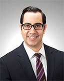
Dr. Williams: And we are so pleased to have Dr. Marcus Malek here with us today from the department of pediatric surgery. Thank you for joining us today.
Dr. Agnihotri: Yeah, thank you.
Dr. Malek: Thanks. Yeah, thanks for having me. It's an honor, and I'm really happy to be here. Thanks guys.
Dr. Williams: So we were chatting just a little bit before we got started recording. And you had said that you were from New Jersey originally. And so I have to ask just one silly question to start with. Is Pittsburgh east coast, or is it Midwest for you as a Jersey boy?
Dr. Malek: Oh, that's a heavy question. We could burn the whole 20 minutes on that, but I don't consider it east coast. I think it's the Northeast.
Dr. Agnihotri: Okay.
Dr. Malek: I don't think it's quite the Midwest. I actually tell people who are thinking of moving here from the New York, New Jersey area that you don't feel too distant from that kind of east coast culture, but you get a little bit of the nice and pleasantries of the Midwest mixed in. So I think it's good. I think it's sort of a mix, yeah.
Dr. Agnihotri: Building on that. So what would be one thing you absolutely love about Pittsburgh?
Dr. Malek: There's a lot. For one thing I met my wife here when I was in the lab back in 2008. And so her family's here and we've started a family here and it's a fantastic place to raise a family. It's a very easy place to live. And of course the healthcare is the thing that brought me here in the first place, and I think we offer tremendous level of care here. And I think that there's so many opportunities. And we're going to talk a little bit about my lab today. And the fact that I have a lab has a lot to do with the environment that I'm in here. Because I've been really a clinician for at least the first five years of my career without any sort of lab work. And it's this kind of environment that allowed it. So, yeah, there's a lot I love about Pittsburgh.
Dr. Williams: Well, you've had an interesting personal story too, with the move and developing your own family here, but let's talk a little bit, like you said, about your career path. How did you end up getting from east coast all the way to Pittsburgh?
Dr. Malek: Oh yeah. It's far. So I've wanted to be a pediatric surgeon since I was a third year medical student. And so it's a pretty tough fellowship to get. And so in order to distinguish yourself, you usually have to do something within your residency beyond just completing your training. So this was the mid-2000s. And now I think a lot of people do various different things, but back then, you pretty much went into a basic science lab.
And so there was really only one pediatric surgeon doing basic science in New York and they were at Columbia and their lab was full. And so I said, okay, I'm open. I didn't have a family. I can go anywhere. And so I started looking for big name researchers in pediatric surgery. And of course my current division chief and boss, George Gittes, his name came up. And so I just emailed him. And George called me probably 10 minutes later and we chatted for a little bit and he said, "come on out." And I went to a couple other places before I came here. And then I came here and I just... First of all, that view when you cross the bridge, the Fort Pitt Tunnel onto the Fort Pitt bridge, that just blew me away.
Dr. Williams: Oh yeah. That's breathtaking.
Dr. Malek: Yeah. Yeah. I was like, "whoa, okay." Okay. This is not what I expected coming from Manhattan. I just didn't know what to expect, but that was a nice sort of good feeling coming into it. And then, of course, when I saw Dr. Gittes's lab, I saw the level of research he was doing and I thought this is definitely the right place for me. So that's what brought me here. And then in the lab, I met my wife. And so in addition to the wonderful medicine and lab opportunities that are here, having her family here and her home base being here made it an easy place for us to settle down in.
Dr. Williams: It sounds like a good career opportunity and nice people to work with too if you met your wife there.
Dr. Malek: Yeah.
Dr. Agnihotri: No for sure. Yeah. Do you want to highlight a little bit more of what you do in terms of surgery and then how that transitioned into your passion for research?
Dr. Malek: Yeah, absolutely. Thanks. So I've always liked surgical oncology, but as I mentioned, I wanted to be a pediatric surgeon and something sort of is happening in pediatric surgery really in the last 10 to 15 years, which is our pediatric surgeons have tended to be sort of the last general surgeons. You know, we do it all. We're in the chest, we're in the neck, we're in the belly. We do everything. That hasn't really persisted in adult surgery. But pediatric surgery is starting to understand that there is so much knowledge being gained year after year within each sort of subspecialty of surgery. That it's good to have somebody that focuses on one thing or another. And so we were starting to do that here. And so I thought, okay, I'd love to really focus on oncology.
But in order to really feel like I was bringing something special or unique to the table, I went to New York to Memorial Sloan Kettering, and I did a pediatric surgical oncology fellowship there, which was fantastic. It allowed me to sort of see things through the eyes of a surgical oncologist, learn to have that sort of mindset and approach, and then bring that back here. So that's what I did when I came back here. And then I noticed when doing cancer surgery in children, we have all this tremendous technology to help us to find the cancers, stage the cancers and things like that. But then you go in the operating room and you're on your own. And we have a lot of training and we're good at it and that's fine.
But sometimes there are small tumors that are difficult to find. And so we just sort of look at the CAT scan and we say, "Okay, it's next to this, it's next to that. Let me look there." But we don't really have anything to help us in the operating room. And further, when we find the tumor, how do you know that you're getting it all out? You just sort of eyeball it and you say, "okay, it looks like...I don't feel more tumor. I don't see any more tumor. I think I got it all out." But I just felt like there's something missing there.
There's another level that we could do. How can we be more certain? What can we do to help us to find these tumors? What can we do to help us to really make sure and confirm that we've gotten the entire tumor out? We've got negative margins, if that's what we're looking for and that sort of thing. And so that has always been on my mind since I started my practice and then the opportunity sort of arose to look into that further. And that's what sparked the creation of my lab.
Dr. Williams: I feel like I'm enamored. I'm like, how do you fix this? Oh my gosh. Because it almost seems archaic at this point in time that we have so much technology and then you go into the operating room and it's like you're back in the 1960s. And it's like, do your thing. You can find it.
Dr. Malek: Yeah.
Dr. Williams: What types of things are you specifically looking at to... I wouldn't say fix the problem, but maybe help the problem in your lab?
Dr. Malek: Yeah. So, I mean, there are already technologies out there that are in use. They're just fairly limited. So sentinel lymph node biopsy is a really good example. So for kids, with melanoma or adults with breast cancer, sentinel lymph node biopsy is standard of care. And it does use imaging or really nuclear medicine technology to help you to find an area of interest. And so that was sort of my first... I've always found that fascinating and I've always been interested in, so how do we take that to the next level. But sentinel lymph node biopsy is really a passive technology. So essentially you're injecting a radio isotope that you can detect with a handheld wand and it gets picked up by lymphatics around your tumor of interest. And then it goes to the draining lymph node. And then you take that lymph node out, assuming that that's the node that drained the tumor since you injected right around the tumor.
So it's very cool. And it's really changed the way that we do those operations. And it's limited morbidity. I mean, about 20% of patients who get an axillary lymphadenectomy end up with lifelong lymphedema. And so that's significant. And that sentinel lymph node biopsy has really reduced that rate significantly.
But it still is a passive technology, right? So if you want to use that in other tumors, it doesn't really work. It doesn't help you to identify another tumor. In this case it's helping you identify the lymph node draining the tumor. So immunotherapy is therapy is really a burgeoning field in oncology, including pediatric oncology. And essentially the concept behind it is you find a tumor-specific antigen or tumor-specific protein or cell surface marker and you have an antibody or some other agent that goes directly to it.
And so they do sometimes just by activating the immune system that in and of itself can create tumor cell death. But sometimes they can, in some cases, they actually attach a chemotherapeutic agent to the antibody and it goes and acts locally. So these things are happening. And I thought, okay, well, why can't we do that, so take an antibody against a tumor specific antigen that ideally there's not a high expression of that antigen in the normal body tissues, so it's very tumor specific, and put something on there that can help me to find it in the operating room? So the technology we already have, we talked about for sentinel lymph node biopsy is a gamma probe, right? So it's a handheld detector that can pick up the gamma rays from a radio isotope.
So you could put a radio isotope on one of these things and use your gamma probe that already exists. The other thing that's really been gaining a lot of enthusiasm is fluorescent guided surgery. And the most common agent used in that is Indocyanine green, which is a near infrared dye. It's been in use since the 1950s or 1960s. So it's decades, it's very safe. We've been using it for a lot of different things. And it's also been used for some tumor surgery, but it's also passive. It tends to accumulate in tumors because the vessels are leaky. They're not normal vessels so that they accumulate in the tumors. And then they don't have good lymphatics so it doesn't really get out of the tumors. So you give ICG, it goes throughout the body, and then it'll sort of collect in the tumors. And then you can use a near infrared camera to visualize it.
But again, it's passive. Areas of inflammation tend to have high ICG signal as well. There's a lot of background signal. It's not going to be good in all tumors. So, while there's a lot of interest in it, and I use it sometimes for various tumors, again, it's not tumor-specific. So similarly, you can add a fluorescent dye to a protein or a mini body or something that can go directly to your antigen of interest. And so you can label it with both a fluorescent dye and a radio isotope, and then you can use your Gamma wand and your neuro infrared camera, and you can specifically see the tumor. So this is the concept behind it, behind all this.
Dr. Agnihotri: So what's fantastic is, I think surgery is one of those rare fields where you really go from bench to bedside. You could play with some of the most amazing toys. I'm always jealous with the cool toys that you have. Can you explain to our audience some of the tumors that you do work on and some of the new techniques that you've been incorporating through your research, anything related to imaging or some of these dyes and what specific tumors you're seeing success or signal in?
Dr. Malek: Yeah, so absolutely. This kind of gets to what's what I think is really special about Pittsburgh is I presented maybe three years ago at our Mellon scholar seminar and the Mellon scholars are the sort of junior faculty or assistant professor level faculty here at CHP.
Dr. Agnihotri: Yeah, that's where I met you.
Dr. Malek: Yeah, you're part of it. And that's where we met. And Gary Kohanbash, who's a researcher here, who's become a good friend, he saw me talking about some of this. And he said he was doing immuno pet for brain tumors and was seeing how that type of work could roll into what I was interested in doing. And he was super excited to have a clinician to work with that could put this stuff into the clinical arena. And so we started talking and we just started building this relationship that has grown ever since. And so at this point, we decided after meeting with our pathologists, our radiologists and our oncologists here, we thought, okay, which tumors could this sort of technology be best for? And the one we came up with, which I, 100% support is neuroblastoma.
And the reason is that neuroblastoma, and in particular high-risk neuroblastoma, it's an awful disease. You know, despite decades of amazing work that's been done, our survival long term is probably only 50% to 60% for high-risk neuroblastoma. And when you talk about surgery for high-risk neuroblastoma, it's one of the most difficult operations that any surgeon will undertake. I mean, these are tumors that wrap around the aorta and all of its branches. And you really want to get at least 90% or, really, I try to get a hundred percent if you can, of the tumor out.
So you can imagine that's a challenge. And in fact, when you go back and you look at the largest databases in Europe, through the SIOPE (The European Society for Paediatric Oncology), which is their kind of their COG (Children’s Oncology Group), where you look at the COG experience here in North America, the rate of inadequate surgery is probably about 30%. Which is, again, easy to understand given how challenging those operations are. And the rate of significant complications in surgery is probably also about 30%, maybe even 40%. So it's sort of the perfect tumor, I think, to use this type of technology for, because it's important to really distinguish, okay, where can I stop? Because it's not like there's a tumor edge that's super clear. I mean, the tissue around it is often thickened and often has some inflammation. These are patients that all receive chemotherapy previously.
So the tumor has died off in certain areas. And so you'll see it's not normal. There's not a very normal transition, but on the other side of tumor, are these vital structures, nerves, vessels that you can't just sacrifice. So this sort of thing would be extremely helpful.
The other thing about neuroblastoma is it tends to spread to regional lymph nodes, and there are often deposits of disease that are distant from the primary tumor that you'll see them on scan, but they can be very hard to find in the operating room. So this sort of technology would lend itself perfectly to neuroblastoma surgery. And I think our review of what's going on in Europe and in North America would show that we need to do better. So neuroblastoma was the first thing to tackle.
So the first question is, okay, do we have an antigen to go after? And we do. These immunotherapy for neuroblastoma already exists. Denintuzumab – It's standard of care for maintenance therapy for high risk neuroblastoma.
So everything just sort of fell into place. We've got an antigen, we have an antibody and we have a disease that could really benefit from this. So we went ahead and we partnered with another really critical person that's helped us to get to the point we're at now, Barry Edwards. Now he is now at University of Missouri, but in today's day and age that's not a boundary. We just hop on Teams or Zoom and we chat and we send him stuff down there. He sends it back up here. But when we first met, he was here, he was at University of Pittsburgh.
So he is a radio chemist essentially. And making these antibodies is easy for him. He's been doing it for decades. And he's been doing it for the most part for imaging purposes and there have been neuroblastoma specific imaging agents based on anti-GD2, but it's never been used to actually guide surgeons in the operating room. So again, he was super excited to join up on this effort as well. And when I asked him, I said, "Oh, I want a fluorescent dye on there. I want a chelator and a radio isotope." He's like, "Oh yeah, that's what I do. That's easy." So we all got together and I mean, we were excited. We thought we could really make an impact and we realized, yeah, we can do this.
Dr. Agnihotri: That's fantastic.
Dr. Williams: So how far along are you in the research process thing? Cause you've talked about the global idea of where you would like to go with this and then it sounds...you just said it's easy. It doesn't sound easy. So how far along are you in this?
Dr. Malek: It didn't sound easy to me either, but Barry promised me it was easy. And Barry was right. Easy enough. Mouse models of neuroblastoma, they exist. And we actually put together a team that has created these mouse models previously. And so we developed our mouse model of neuroblastoma here, and it was up and running pretty quickly. And we actually... put the tumors directly in the adrenal gland. So it's called an orthotopic model. So it's exactly where the tumor ought to be. And I think that's important for this type of study. You could put them subcutaneously and they're super obvious and it's easy to study, but it's more interesting to be able to go in and find it and differentiate it from normal tissues. So we developed the mouse model and sure enough, we gave Barry some anti-GD2 and he made the dual labeled antibody.
Dr. Williams: What a magic man.
Dr. Malek: Yeah, right. And then of course, does it work? Right? So we went ahead, we injected it in the mice and it works. There is strong gamma signal in the tumors. You can differentiate it very easily from normal mouse tissues and the tumors fluoresce. And when we ran a biodistribution, which is basically you take various tissues throughout the body, and you check for levels of antibody. There was a clear spike in the tumor. There's a little spike in liver because antibodies tend to end up in the liver and a little spike in the heart because there's a lot of blood in there. But in general, I mean, the signal for the tumor was at least three times greater than anywhere else. So we had our antibody.
Dr. Agnihotri: That's super exciting. Can you convey to our audience...because this is amazing work. Once you see positive signal, safety in a mouse, the next step would be eventually to get into patients.
Dr. Malek: Yeah.
Dr. Agnihotri: So what's your vision or your long term plan for that? Because this is fantastic.
Dr. Malek: Yeah. You're exactly right, Sameer. That's what we're talking about now. So we've already started to talk with our clinical trials and translational folks here at Pitt. And that is our aim. Obviously we need to ensure safety and learn about what's called the "pharm tox." Right. So the toxicology and what are the pharmacokinetics of this a little bit greater before we can go into humans, but that's the next step is to run those studies and get a set-up for a clinical trial. I'm hoping we can be in patients within the next year or two.
Dr. Agnihotri: Wow.
Dr. Williams: Yeah. That's truly amazing. And I know that you're doing this with your department of pediatric surgery as well, since we're almost at the end of our 20 minutes, which I feel like just flew by during this episode.
Dr. Malek: Yeah.
Dr. Williams: Do you have any social media platforms that you use to talk more about this research or any other handles that you'd like to tell people about in case they wanted to look up more information?
Dr. Malek: Yeah. So I try on Twitter. I'm not a Twitter guru by any means, but I am on Twitter. My handle is @drmarcusmalek but I don't necessarily use that to talk much about research. I try to just be aware of what's going on. My research is probably more so you'd find it on my website through the Mellon Institute. So you can find it there. If you go to the children's page and you look at the research tab, you can find my lab there and it's got pretty up-to-date information on there, including all the members of the lab who've made this all happen.
Dr. Williams: Perfect. Well, this is amazing. Thank you so much again for coming today and talking with us, this was truly interesting and novel research that we were excited to hear about.
Dr. Malek: Yeah, thanks for having me. You're right, it went by fast. It's always exciting to talk about this and I appreciate the opportunity to be here. Thanks.
Dr. Williams: And thank you all for listening to "That's Pediatrics."
Voiceover: You can find other episodes of "That's Pediatrics" on Apple Podcasts, Google Podcasts, Spotify, and YouTube. For more information about this podcast or our guests, please visit chp.edu/thatspediatrics. If you've enjoyed this episode, please be sure to rate, review and subscribe to keep up with our new content. You can also email us at podcast.upmc@gmail.com with any feedback or ideas for topics you'd like our experts to cover on future episodes. Thank you again for listening to "That's Pediatrics." Tune in next time.


